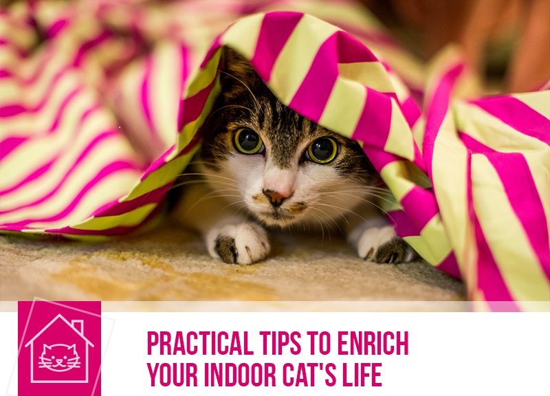
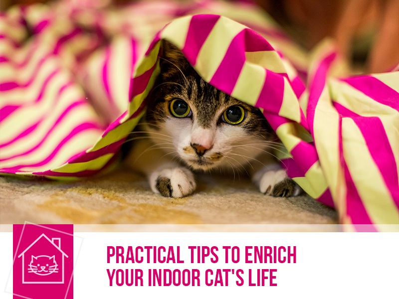
Diabetes mellitus in cats

cats-33-diabetes-mellitus-in-cats

Many owners will be familiar with diabetes mellitus, as it’s an unfortunately common disorder in humans and there have been many public health initiatives towards making diabetes a known issue.

Diabetes mellitus is an endocrine disorder that influences the mechanism that regulates sugar concentration in blood. Our organism’s capacity to maintain a relatively stable concentration of glucose circulating in the blood stream is one of the most important homeostatic functions. Glucose is the chief energy source of our body and any extreme fluctuations, either too little or too much, will be extremely harmful.

Insulin is the hormone responsible for reducing the excess blood sugar to normal levels and making glucose available as an energy source. It is produced in the pancreas by secretory cells called β-cells and in the most common form of diabetes in cats there is a progressive destruction of these cells.

There are several clinical signs to look out for in our furry friends: increased urine production, increased water intake, increased appetite and unexplained weight loss. Try to keep an eye on their behaviour too. Diabetic cats might be more lethargic, interact less with the family and not be able to do as much grooming, so they end up looking rather unkempt.

If all the signs mentioned above are missed or devalued, it might evolve to include neuropathy of the pelvic limbs (limb weakness, ataxia and incapacity to jump) and diabetic ketoacidosis.

Diabetic ketoacidosis is a serious complication of untreated diabetes or of incorrectly supplemented insulin therapies. This causes many ill effects and is a medical emergency, so if you see vomit, lethargy, depression, high respiratory rate, weight loss, icterus or lowered body temperature in a diabetic cat or a cat that may have diabetes, don’t hesitate in bringing them in to the vet!

Unfortunately, diabetes mellitus is often a chronic disease that has no cure however, it can be medically controlled and your cat can have a long, healthy life. It’s important that the earliest diagnostic possible is made to begin a successful management of insulin, sugar levels and not risk further complications.

If you think you cat might have diabetes mellitus, take them to the vet and share your concerns. Even after a successful diagnosis and therapy, it’s important to check in with your vet frequently to make sure everything is still running smoothly.

Would you like to know more about cats? Check our Feline Courses:

Feline courses

Importance of adequate nutrition for orphaned or sick foals

horse-43-importance-of-adequate-nutrition-for-orphaned-or-sick-foals

Foals of different ages will have differing nutritional requirements. As such, the proper dietary composition will change as the foal grows. This is especially important as they develop from a newborn to a weanling.

The transition from being fed exclusively via the placenta, with maternal energy sources to the foal having to procure their own energy is a stressful metabolic transition. Normal neonatal (newborn) foals are at risk of hypoglycaemia, due to high metabolic rates and transition from womb life to outside life, and sick neonates are even more at risk of falling into severe hypoglycaemia. Hypoglycaemia, also called low blood sugar, can cause weakness, abnormal gait, collapse, intolerance to movement and if severe and unresolved, it might lead to seizures or death.

Foals, and baby animals in general (like lambs or calves), are particularly sensitive to adverse conditions like hunger and cold because they do not have energy or fat stores to draw upon in time of stress. Added to that, they have immense growth in a relatively short amount of time. A newborn foal of a light breed will gain in excess of 1 to 1.5 kg of body mass per day. In their first few days of life, a baby horse will consume from 15% to 25% of their bodyweight in milk, or about 8 to 15 litres of milk. Foals will begin to consume and adequately process solids within several weeks of life via a gradual transition from a milk exclusive diet to a forage based diet.

As such, an adequate and proper nutrition is paramount in dealing with abnormal situations such as an orphan or sick foal.

There are many ways to rear these animals into amazing, healthy adults but it’s something that requires solid knowledge of the horse’s needs and a strong commitment to make sure things like hypoglycaemia or digestive issues won’t hurt your animal. This is a situation that requires the expertise and attention of a vet, so if you find yourself in hands with a young horse with special needs don’t hesitate to contact your clinician and they will advise you on how to best proceed and help you create a feeding plan that fits their needs.

If you would like to know more about equine nutrition have a look at our course at: https://www.thepetprofessionals.co.uk/courses/product.php?u=P5T7PJ9EAJBCEAKZTKJ4XMRH

Would you like to know more about horses? Check our Equine Courses:

Equine courses

Hypertrophic cardiomyopathy in cats

cats-32-hypertrophic-cardiomyopathy-in-cats

Having a dodgy ticker isn’t just limited to people, much to our feline friends’ chagrin they can suffer from heart problems pretty much in the same way.

Hypertrophic cardiomyopathy is a rather big, mean looking word for a relatively common heart problem in our kitties. Hypertrophic means that the muscle that composes the heart is much larger than what it should be and cardiomyopathy means the problem is in the heart muscle.

It’s not known why the heart walls thicken so dramatically but some breeds like Maine Coon and Ragdoll have mutations in a gene that codes for a cardiac protein. This dysfunction is implicated in hypertrophic cardiomyopathy, however, a large majority of the cases we don’t know the cause.

It manifests at a mean age of 5 to 7 years with a larger predominance towards male cats.

The clinical signs that should be looked out for are difficulty breathing, not wanting to move much, vomiting, not wanting to eat, fainting (called a syncope) and if it progresses enough there might be sudden death due to heart failure. Discerning owners that also have dogs might know that that the usual cause of doggy heart issues causes cough (because of fluid build-up in the lungs secondary to heart failure) but cats usually do not have cough due to heart problems.

It sounds a bit of a contradiction, if the heart has more muscle it should work better, not worse. While at first, there might be more powerful contractions and better blood ejection to the whole body, the excess muscle soon starts failing. What’s worse is the chamber affected is the left ventricle. This chamber receives a large quantity of oxygenated blood from the left atrium and distributes it towards all tissues and the muscle will occupy a significant portion of it. All problems stem from this lack of chamber volume and the progressive weakening of the muscles because it’s harder for the blood to enter it, so a huge spike in blood pressure in the lungs happens and organs will have insufficient blood supply.

Unfortunately, while it can be managed to an extent it cannot be cured and cats afflicted with this and showing clinical signs have a reduced life expectancy.

Keep an eye on your cat’s tick tacking heart and if you see any of the above, take them in to your vet’s for a check-up. The earlier its diagnosed, the better it can be managed.

Would you like to know more about cats? Check our Feline Courses:

Feline courses

Bloat or gastric dilation in dogs

dogs-49-bloat-or-gastric-dilation-in-dogs

Gastric dilation or bloat is a condition that all dog owners should be familiar with, since it can be deadly if it is not diagnosed and treated on time.

Dogs affected by this condition present a distended stomach filled with gas and/or fluid, rotated along its short axis. This will lead to pain, cardiac arrhythmias, respiratory distress, shock and eventually death.

Owners that have seen their dog suffer from this condition often describe signs such as anxious behaviour, abdominal pain, with pets often pacing trying to find some comfort, unproductive retching, vomiting thick saliva, an elevated respiratory rate, sometimes visible abdominal distension, and collapse.

Although the mechanism involved is not entirely understood, it is thought that this condition can be caused and influenced by several factors. The chances of a dog suffering from bloat seem to be affected by genetics as well as environmental factors.

This condition is more commonly seen in large or giant breeds of dogs such as the German Shepherd, the Standard Poodle, the Great Dane, and the Irish Setter, among others. Dogs that are older, deep-chested, and with abnormal gastric motility are at a higher risk of gastric dilation. Bloat is very uncommon in small dogs and in cats.

If you notice any of the previously mentioned signs on your dog, do not hesitate and get him or her to the vet! Quick gastric decompression is key for the dog’s survival and to reduce the chances of further complications.

The recommendations to prevent this problem include feeding your canine companions with two to three smaller meals a day instead of offering just one large meal and avoid using an elevated platform to place their bowl. There was a time where it was thought that using a bowl normally placed on the floor might encourage dogs more susceptible to bloat to swallow more air (aerophagia). However, over time, it has been shown that feeding your dog from an elevated platform is also a factor that predisposes to the occurrence of this condition.

Would you like to know more about dogs? Check our Canine Courses:

Canine courses

Feline Leukemia Virus (FeLv) in cats

cats-31-feline-leukemia-virus-felv-in-cats

If you are a cat owner you have probably heard of this disease before. The Feline Leukemia Virus (FeLV) is a retrovirus that affects cats all over the world.

Our feline companions become infected through close contact, primarily through saliva. The virus is passed among cats that spend a lot of time together, as they share food and water bowls, use the same litter boxes and often groom each other. Kittens can be infected through their mother’s placenta or when she licks and nurses them. Entire male cats allowed to roam are also at a higher risk of becoming infected since these cats often fight and bite each other.

Once a cat is infected, the virus starts replicating in the respiratory epithelium, salivary glands and it can replicate in the bone marrow. The outcome of this disease depends on the age, immune status and genetics of the cats involved as well as the pathogenicity of the virus and virus concentration. In general, young and immunosuppressed cats have a higher probability of developing a progressive infection.

Cats that develop a progressive infection can present several FeLV-related disorders such as anaemia, immunosuppression (cats become more susceptible to secondary infections), immune-mediated diseases, stomatitis, and reproductive, digestive and neurologic problems. This virus is also capable of oncogenesis, which means it can be responsible for the appearance of malignant tumours such as lymphoma, leukemia, and other hematopoietic tumours.

Although this condition can be fatal, with cats succumbing about three years following their diagnosis, other cats can stay healthy for many years. Survival rates are also much higher for single cats, living indoors that receive appropriate veterinary assistance (flea/tick treatment, worming, vaccination, neutering, etc.).

Cats diagnosed with FeLV should be fed a good quality diet (avoid uncooked food and unpasteurized milk due to the risk of bacterial and parasitic infections), visit the vet frequently for check-ups and it is also recommended to keep these cats indoors to avoid further transmission of the virus.

The Feline Leukemia Virus is very fragile, being susceptible to soaps, disinfectants and variations in temperature. It will become inactivated in the environment within minutes and this is why very close contact is necessary for transmission.

The prevalence of this disease has decreased over time, possibly due to more effective control measures and to the increased number of cats being vaccinated. It is important to keep in mind that the vaccine does not guarantee 100% protection against progressive infection, but is has been an important factor in reducing the prevalence of this disease.

Testing for FeLV and applying measures to prevent healthy cats from being exposed to this virus is the most effective way to prevent this infection from spreading. It is recommended for owners to get their cats tested. This infection has very serious health consequences not only for infected cats but also for healthy cats.

Would you like to know more about cats? Check our Feline Courses:

Feline courses

Babesiosis in horses

horse-42-babesiosis-in-horses

Babesiosis, also known as equine pyroplasmosis, is a non-contagious tick-born disease caused by the Babesia caballi and Babesia equi especies of protozoa.

This infection is a problem in international horse trading and competitions. Many countries, like the United States, will have import regulations regarding the infection status. Animals that never have been infected that come in contact with infected ticks will most likely develop the disease.

Ticks house these parasites and transmit them to the horse when feeding but it cannot be passed from horse to horse, unless there is blood transfer between animals (like on needles, blood transfusions or any sharp instrument with contaminated blood). Usually though, infected horses act as the source of the infection for ticks and then the ticks will go on to infect other animals.

It causes severe destruction of red blood cells and in the case of B.caballi, the parasite itself might clog the smaller calibre vessels in the lungs, liver or even central nervous system, damaging them.

With the progressive destruction of red blood cells and the increase in parasite numbers, affected animals will display reluctance to exercise, fever, anorexia, small dotting haemorrhages in mucous membranes (like inside the mouth, vulva or eye), dehydration, colic, abdomen 0r limb oedema and/or difficulty breathing.

These clinical signs usually only manifest in acute diseases. Animals that manage to survive this may become persistent yet immune carriers and may generate chances for ticks to infect animals that have never contacted babesiosis.

The best way to prevent babesios is to run a tight ship regarding tick control on your horses and always follow good biosecurity measures (no reusing needles, always disinfecting sharp instruments between horses).

Would you like to know more about horses? Check our Equine Courses:

Equine courses

Notoedric Mange


cats-30-notoedric-mange

Notoedric mange is a skin problem of cats caused by Notoedres cati, one of the species of mites that causes mange in pets. This problem may affect any cat, regardless of age or gender and, while it’s fairly specific for cats, it might affect dogs and rabbits as well.

It’s a parasite transmitted by directed contact and is especially important in places where several cats are gathered like colonies (both domestic and feral), hotels, veterinary clinics or even multi cat homes. When a cat starts manifesting clinical signs, all other cats in contact will, usually, be affected as well.

Important clinical signs to look out for include:

– Intense itch that does not seem to be related to any season or weather condition;

– Itching that might start out mild but will progress to severe and distressing;

– Skin crusts and lack of fur that start on the ears and then spreads to their face, hands and feet, and eventually with the whole body being affected;

– Incessant scratching and grooming will eventually lead to severe self-trauma.

Due to animals scratching and traumatizing their own skin, this will be fertile breeding ground for skin infections because intact skin is a very important barrier to bacteria. If this condition progresses, treatment for both the mites and the infection that had the opportunity to develop will be required so prompt action will save you and your cat much trouble.

If you notice any of these clinical signs on your cat consult with your veterinary surgeon. The treatment for this mange should be administered on all cats that are in contact with each other and it lasts for several weeks. If the chosen treatment involves dips of insecticides it is very important to ensure the product covers all of their skin, including head and ears otherwise the treatment will most likely fail and these cats will re-infect other cats that might have already recovered. If skin infections have already developed, a course of antibiotics will be required to fully kick away this disease.

The good news is, if all animals are correctly treated symptoms will be significantly reduced within 2 weeks.

It’s important to keep in mind that notoedric mange is a zoonosis, meaning that this mite will infect humans and cause itch and rashes on arms, chest and abdomen of those who have been in closer contact with infected cats. Fortunately, it’s usually solved spontaneously in about 10 days after all animals have been treated.

Be on the lookout for this bothersome problem in our feline friends to catch it early on!

Would you like to know more about cats? Check our Feline Courses:

Feline courses

Abnormal increase of volume in the scrotum

horse-41-abnormal-increase-of-volume-in-the-scrotum

The wellness of external genitalia is a very important component of the reproductive health in horses, especially in male horses. The scrotum houses the testicles and all the vascular and nerve support for them, functioning as one of the linchpins of the horse’s reproductive system.

It is good practice to pay close attention and know scrotal characteristics, often how fast there is veterinary intervention will determine the outcome and future reproductive capacities of the stallion. Fortunately, it’s an area that sits between the horse’s hind legs and is reasonably well protected from harm.

Be on the lookout not only on size changes but also position and the presence of wounds on the skin or deeper tissues. The stallion’s behaviour may be changed due to pain, they may become reluctant to jumping and even walking.

The causes of an abnormal increase of size are plentiful. The most common causes are trauma, scrotal or inguinal hernias (passing of other abdominal organs into the scrotum e.g.: small intestine lodged in scrotum due to weak muscles/rupture in the abdomen) and testicular torsion.

Other important causes include cancer, bacterial, viral or parasite infections or even auto-immune disorders (the horse’s immune system is actively targeting its own tissues, usually the testicles and damaging them.)

Some of these conditions can cause irreversible impairment of fertility, especially if left without proper veterinary care for long periods of time. Semen collection and evaluation should be performed at around 90 days after the cause has been resolved or after surgery.

Keep an eye out on your stallion and save you (and him!) a world of trouble!

Would you like to know more about horses? Check our Equine Courses:

Equine courses

Equine eye conditions

Horse 40 - Equine eye conditions

Horses suffering from an eye condition may display several signs of discomfort such as eye rubbing, excessive blinking, an increase in the amount of tears, an abnormal shape or size of one or both eyes, changes in the cornea, drooping of the upper eyelid, abnormal discharges, being more sensitive to light, pain, holding the eye shut and may even become depressed.

The list of common eye problems in horses includes uveitis, ulcers, cataracts, tumours traumatic injuries, and foreign bodies, among others.

Uveitis
Uveitis is the term used for an inflammation involving different structures inside the eye that can have many different causes such as bacteria, viruses, neoplasia or even trauma. This is a painful condition that over time can lead to the formation of adhesions within the eye and eventually lead to loss of vision in the affected eye. Uveitis is a common cause of blindness in horses and in some cases the affected eye may be surgically removed.

Equine recurrent uveitis (ERU) is a complex immune-mediated disease that can be more or less serious depending on the severity of the uveitis, duration of episodes and response to treatment. Prompt veterinary assistance is key since the sooner the inflammation is under control the better. This will limit the damage to the structures inside of the eye.

Corneal Ulcers
A corneal ulcer is an open sore in the transparent membrane that covers the eye (the cornea) which is caused, in most cases, by an initial trauma. Corneal ulcers also cause inflammation, pain and there is a risk for secondary infections.

Cataracts
A cataract is an opacity (cloudiness) in the lens of the eye, a firm, biconvex structure filled with gel that is suspended behind the iris. Cataracts can be congenital or can be acquired throughout the animal’s life. Common causes include trauma, nutritional abnormalities, toxicities, inflammation or be secondary to other conditions such as ERU. Older horses can also present cataracts (senile cataracts) since this condition is a normal part of the normal ageing process of the lens.

Tumours
There are several types of tumours that can affect the eye itself and surrounding structures such as sarcoid, melanoma and squamous cell carcinoma.

Foreign bodies
Despite the eyes’ blink response which is a natural mechanism that protects the eye from external aggression, a foreign body can go into the eye. The most common foreign bodies affecting a horse’s eye include twigs, thorns, grass seeds, and these need to be removed as soon as possible.

It is very important to take your horse to the vet as soon as you realise there is something unusual with your horse’s eyes because what can start as being a minor problem can become more serious. Please keep in mind that many eye issues also cause a lot of discomfort and pain. Eyelid wounds, painful eyes, foreign bodies, all these conditions are regarded as emergencies, so it is recommended to get your horse checked by a vet straight away.

Would you like to know more about horses? Check our Equine Courses:

Equine courses

Feline Lower Urinary Tract Disease

Cats 29 - Feline Lower Urinary Tract Disease

Feline Lower Urinary Tract Disease (FLUTD) is a term used to describe conditions that can affect the lower urinary tract of cats (the bladder and/or the urethra – the tube that carries urine from the bladder to the outside of the body). The conditions which cause it can vary, but the signs are generally very similar.

Cats with FLUTD typically have difficulties urinating (crying and yowling with pain when urinating) and urinate more times than usual. They may also have blood in their urine and urinate outside the litter box. Affected cats may only be able to pass small amounts of urine or no urine at all. Please keep in mind that cats unable to pass any urine at all should been taken to vet immediately, as this is an emergency!

FLUTD occurs most frequently in middle aged, overweight cats that get little exercise, use an indoor litter box, have restricted access outside and eat a dry diet, but it can occur in any cat.  Stress may also increase the risk of a cat suffering from this condition.

Urinary tract infections, urinary stones and feline idiopathic cystitis are amongst the most common conditions that can affect the bladder and urethra of cats. Feline idiopathic cystitis is a disease which the cause is unknown. It involves inflammation of the bladder and it is thought stress plays a major role in its development.

In some cases, a more severe condition can occur: the development of a blockage in the urethra. This prevents the cat from passing urine and emptying the bladder. This condition occurs most frequently in male cats and if left untreated it can be fatal.

Feline Lower Urinary Tract Disease has different treatment approaches, so if you notice any of these signs on your cat do not hesitate and take your pet to the vet!

Would you like to know more about cats? Check our Feline Courses:

Feline courses

Just another WordPress site