Interpreting pain using facial expressions

Detecting pain responses in our faithful companions as soon as possible is one of the most fundamental ways to ensure that illness is investigated and treated in a timely fashion.

However, detecting pain in animals, especially horses that are being ridden, can sometimes be quite complicated. Research has shown that there is a poor interpretation of pain and lameness in all people involved in horse training. This has the obvious issue that it delays treatment and means that treatable conditions are potentially only detected when treatment is either impossible or much harder.

To simplify identification of pain in horses, research has looked into a promising new direction: facial pain expressions. By looking at how animals react to pain, it may be possible to develop assessment protocols that everyone can use and interpret.

There are certain facial features that might indicate underlying pain such as ears pinned back, eyelids that are partially or completely closed, tension in the muscles above the eye, straining mouth and pronounced chin, strained nostrils and prominent chewing muscles.

Investigation into area has led to the creation of several pain assessment scales with the ultimate goal of making pain assessment as accessible and as accurate as possible, with exciting new developments on the horizon!

Would you like to know more about horses? Check our Equine Courses:

Equine courses

Recognising osteoarthritis in cats: What difference does it make?


Loving owners want to see their cats happily purring all day long! Unfortunately, many cats struggle with pain and discomfort due to an illness that for a whole host of reasons is not easy to see.

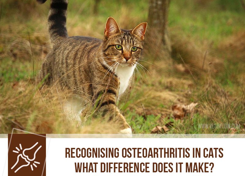
Osteoarthritis is, to put it simply, a long-standing inflammation that affects the different components and tissues of a joint (usually the elbows, knees and hips). As time goes by, the joint becomes damaged by this ongoing process, causing persistent and long-lasting pain.

This process of deterioration, also known as degenerative joint disease, can begin due to joint abnormalities, trauma, and of course, long-term use, but in most cases, there isn’t a single cause or event we can point to as being the culprit. We know that it affects middle-aged to older cats and that this disease is severely underdiagnosed in our feline friends. There are two main reasons for this.

First of all, cats struggling with the condition will find ways to adapt and cope with their aches and pains, mastering the art of hiding any mobility issues. Arthritic cats may show some stiffness when trying to get out of their beds and in general reduce their levels of activity, spending more time resting or sleeping.

They also start avoiding movements that may be more painful like jumping. This will often result in sleeping in new locations, as well as changes in their toileting habits. Affected cats may avoid using the litter tray as they find it harder to get into the tray. This leads to house soiling and other abnormal toileting behaviours.

Cats with chronic joint pain show other significant changes like overgrown nails and a scruffy or matted coat, as it becomes harder to groom. Changes in character are also common such as being more distant, more vocal, and even becoming aggressive.

Now this is the time to mention the second reason why osteoarthritis often slips under the radar. All these changes (if they are noticed) are often seen as something to be expected from an older cat and not as manifestations of a condition that needs to be addressed.

There are many options to consider: weight loss to reduce unnecessary strain on affected joints, medication and acupuncture to help with pain relief, and physiotherapy.

Simple environmental changes such as enabling easy access to all important resources such as food, water, and providing low-sided litter trays can make a huge difference in the life of an arthritic cat. Ramps or steps can help reach higher levels, well-padded beds offer extra comfort and a helping hand with grooming will be greatly appreciated.

Unfortunately, osteoarthritis has no cure but there is plenty that can be done to improve quality of life, reduce the possibility of further joint damage, and most importantly, to bring much needed comfort to cats struggling to cope.

If you’re concerned that your cat may be affected by osteoarthritis, schedule an appointment with your vet to discuss treatment options!

Would you like to know more about cats? Check our Feline Courses:

Feline courses

Cryptorchidism

Cryptorchidism is described as the failure of one or both testicles to descend to the scrotum. This descent usually happens in about 2 to 6 months as animals are born with testicles that reside in their abdomen and then slowly but surely descend to their final position.

As far as we know, what causes testicles to not descend correctly is most likely genetic but there isn’t much information available in cats. It’s much more frequent and most often talked about in dogs, especially because there are several dog breeds who have a higher incidence – for cats there is about a 1 to 1.7% incidence overall.

They can be palpated if they are retained in the inguinal canal but if they are still in the abdomen, advanced imaging like an ultrasound will be required to see if they are really there or if for some reason they failed to form at all.

This distinction is particularly important because, while a retained testicle by itself is not painful, these abdominal or inguinal testicles are at a much higher risk of cancer or of twisting the blood vessels, nerves and muscles that support them. Be on the lookout for acute abdominal pain in your cat, as this might indicate a torsion has happened and needs to be seen by a vet!

Cats that only have one retained testicle may still be fertile but as it is a genetic condition, it’s best to remove these cats from the breeding pool to lower the risk of other animals with retained testicles.

Do you suspect your purring partner may have cryptorchidism? Bring them over to your vet. They will be more than happy to do the necessary tests to confirm what is going on and recommend the best way to proceed!

Would you like to know more about cats and dogs? Check our Feline and Canine Courses:

Feline courses

Canine courses

Blood in a dog’s urine

Spotting blood in a dog’s pee is usually an alarming finding for pet owners. This is definitely a sign that should not be ignored but there are several conditions that can cause the appearance of blood in a dog’s urine. More than often the reason behind it can be treated.

The presence of an abnormally high number of red blood cells in your pet’s urine causing it to turn from a clear or yellow colour to a reddish-pink colour, is also called haematuria. In some cases, haematuria is not visible to the naked eye and may only be detected when your pet’s urine is examined through a microscope.

The list for the most common causes of haematuria is long but it typically includes urinary tract infections, the formation of urinary crystals and sometimes of bladders stones (urolithiasis), prostatic disease, trauma, and urinary tumours.

Bacterial urinary tract infections are common in our canine friends (particularly in females) and pooches struggling with recurrent bacterial infections have a higher chance of developing urinary stones or uroliths. These are literally rocks that are formed within our pet’s urinary tract. These uroliths can block a dog’s urethra, causing urinating difficulties, and if they become big enough, they can even be palpated through a dog’s abdomen.

Prostatic disease in males includes a wide range of issues such as inflammation (prostatitis), abscesses, cysts, and neoplasia (benign or malignant). All tumours located in the kidneys, ureters, bladder and urethra can cause the appearance of blood in urine.

It is very important to take your pet to the vet for a thorough investigation of the cause or causes of haematuria. It’s not uncommon for a dog to have more than one issue causing these clinical signs.

Although it may not be the most exciting aspect of our dogs’ lives (for us humans, at least), keeping an eye on our dogs’ urine is one of many ways to monitor their health. Any changes in frequency, quantity, colour, and detecting any kind of discomfort while urinating can be an indicator of a potential health issue.

Would you like to know more about dogs? Check our Canine Courses:

Canine courses

Anaplasmosis in cats

The temperature is rising, the days are longer and the sun is actually around! Summer is one of the best times of the year for many of us but with all of these good things also come some particular dangers.

Ticks are parasites that consume our pets’ blood and in exchange, they drop nasty microorganisms in their bloodstream. A lovely deal!

There are many diseases that can be transmitted by ticks and one of them is called anaplamosis caused by Anaplasma phagocytophilum in cats. This bacterium is from the Rickettsiales order which includes other important blood parasites like Ehrlichia and Rickettsia. It has a pretty vast geographic distribution because its tick vectors are found through a wide area and climate, so it’s definitely something that you can stumble upon.

After a tick bite, the bacteria are inoculated in the blood stream where they will infect white blood cells and replicate, killing the original cell and moving on to infect other cells.

Infected cats might present with non-specific signs like fever, anorexia, lethargy and dehydration and sometimes lameness, swollen joints, nose bleeding and abdominal pain have been reported. The course is generally mild and can be resolved quickly, especially with a little help from friendly antibiotics which is good news for our fuzzy buddies.

It’s important to keep in mind that prevention is the best way to make sure nothing bad happens so staying on top of flea and tick treatment is paramount! Talk to your vet about the best way to control these nasty bugs that best fits your pet and their lifestyle.

Would you like to know more about cats? Check our Feline Courses:

Feline courses

Gastric ulcers

Ulcers! The medical term for an organ or body surface that had a chunk torn off of it due to a damaging influence.

In this case, gastric ulcers are when the surface of the horse’s stomach has been eroded and there have been damages to this organ. This, understandably, is quite a problem.

The equine stomach is essentially composed of two parts, a part that secrets several compounds including stomach acid called the glandular mucosa and a part that doesn’t produce any compounds called the squamous mucosa. In between the two there is also a border structure called the margo plicatus that is also not glandular.

Gastric ulcers happen as a part of a disease called Equine Gastric Ulcer Syndrome or EGUS for short. Despite being a very common occurrence, the reason for why the ulcers happen is still under study but we know it to be a complex interaction of situations in the horse.

Both foals and adult horses can have gastric ulcers, though they tend to present differently and affect different areas of the stomach. In foals, there is a tendency to primarily affect the glandular portion while adults suffer more damages in the non-glandular portion.

What to expect from adults affected with EGUS? Unfortunately, nothing too specific. You may see your horse not eating as much, poor body condition, loss of weight, mild or recurrent colic, loose faeces and poor performance.

There are several risk factors that were identified, like stress, transportation, high-energy feed and intense exercise. Sports horses are particularly at risk of ulcers, they have a chance of having them that is nearing 100% because of how they are exercised.

Do you feel your horse might be having issues with ulcers? Contact your vet and they will be more than happy to help you out to diagnose and then manage this condition.

Would you like to know more about horses? Check our Equine Courses:

Equine courses